Microscopy
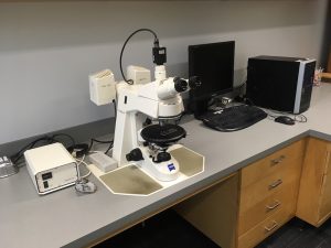
One of the first steps in most geochemical studies (after mapping and sampling) is petrographic characterization of the rocks of interest through hand sample description and microscopy. Transmitted, reflected, infrared and ultraviolet fluorescence microscopy are available at our laboratory. Many ore minerals are opaque. We can use infrared microscopy to see through some of those opaque minerals in order to identify phases that were trapped during ore mineral growth. Ultraviolet fluorescence microscopy is particularly efficient at exciting organics such as hydrocarbons or organic matter. We are equipped with a Zeiss Axioskop 40 Pol with standard transmitted, HAL100 (reflected) and HBO50 (ultraviolet) illuminators and coupled to a FLIR Grasshopper camera.
Thermometry
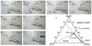
Several thermobarometric techniques (e.g., use of mineral, fluid and melt inclusions) and experimental techniques (e.g., speciation in solvents as a function of temperature) require observing phase changes or in-situ measures at variable temperatures. A USGS gas flow stage, and Linkam stages THMS600 (-196 to 600°C) and TS1200 (ambient to 1200°C) coupled to an Olympus BX53 microscope are shared with Matthew Steele-MacInnis.
Raman spectroscopy
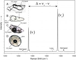
Raman spectroscopy provides information on how atoms interact with each other in minerals and fluids. Because this interaction is dependent on pressure, temperature and phase composition, Raman can be used to interpret some of these parameters. Some applications of Raman include mineral identification (of minerals exposed at the surface as well as included in other minerals), interpretation of how metals are transported in solution, or determination of the pressure and temperature of inclusions trapped in other minerals.
Chemical laboratory
Much of our work requires delicate sample preparation and experimental work. We are equipped with a number of hot-stirring isotemp plates, sonic stirrers, and a vacuum oven for chemical work. In addition, we are equipped with a Zeiss stereoscope, and a Buehler precision saw for preparation of delicate natural samples and experimental loads. Sample slabbing, thin section and mound preparation facilities are also available through the Earth and Atmospheric Sciences department.
Ion Microprobe (SIMS)
We work with Richard Stern at the Canadian Center for Isotopic Microanalysis supervised by Thomas Stachel towards understanding the evolution of fluid sources in ore deposits. Stable isotope geochemistry provides information on the temperature and potential sources of fluids responsible for mineral precipitation. Because minerals can grow over long periods of time, the element sources or temperatures can vary during mineral growth providing a microscopic record of geologic-time processes.
Imaging and point spectroscopy (shortwave and longwave)
We work with Jilu Feng at the core sensing lab led by Benoit Rivard towards research on alteration mineralogy. Clay mineral identification is critical to characterize alteration mineralogy. However, hand sample and microscope identification of clay minerals is often ambiguous. Hydrous minerals absorb natural light at different short wave infrared and near visible wavelengths depending on how elements (and particularly hydrous groups) are bonded within the mineral. Spectroscopy can be applied at a variety of scales from hyperspectral (fly by), to core scanning with resolutions down to 60 micron. Because the bonds present in different clay minerals differ from each other, spectroscopy provides semiquantitative information on mineralogy.
Scanning electron microscope cathodoluminescence (SEM-CL)
We work with Nathan Gerein at the Scanning Electron Microscope Laboratory towards determining the relationships between different minerals in ore deposits. Textural information is critical to determine when different phases precipitate in an ore deposit relative to each other. This textural information is commonly invisible in microscopy but can be shown in SEM-CL.
Additional facilities
The facilities listed above are being used in ongoing projects. Additional facilities that we plan to use in these and other upcoming projects include laser ablation inductively coupled mass spectroscopy through the Arctic resources laboratory lead by Graham Pearson and the ICPMS facility run by Andy Dufresne, electron microprobe run by Andrew Locock, and the micro-XRF laboratory at nanoFAB run by Peng Li.